

Contacts and Location
| Contact 1 | Mārtiņš Borodušķis |
|---|---|
| Enquire about this equipment | |
| Organisational Unit |
University of Latvia Faculty of Biology Chair of Microbiology and Biotechnology - Latvia, Rīga |
| Building | Rīga, Jelgavas 1 |
Description
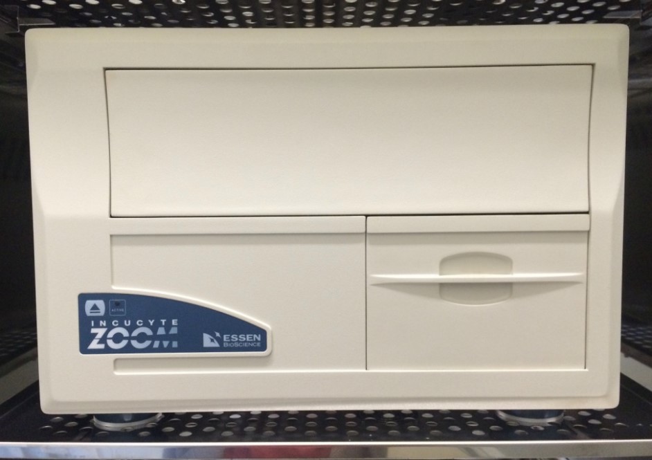
IncuCyte® ZOOM System enables observation and quantification of cell behavior over time by automatically gathering and analyzing images around the clock within a standard incubator
Specification
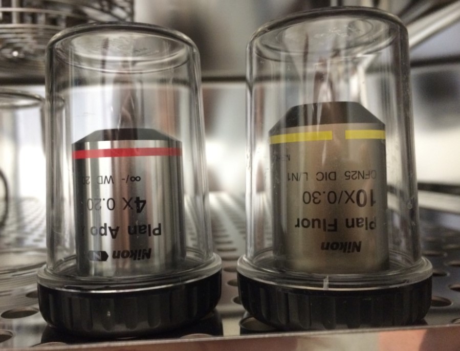
• IncuCyte® ZOOM System consists of a microscope gantry that resides in the cell incubator, and a networked external controller hard drive that gathers and processes image data • Different microscope objectives (4x, 10x, 20x) • Houses multiple T-flasks or microtiter plates (up to 6) and can acquire >2000 images per hour • Supports over 300 different standard plates (including 96- and 384-well), T-flasks, dishes and microslides • Automated time-lapse imaging of multiple positions with 4x (0.2 NA), 10x (0.3 NA) or 20x (0.45 NA) lenses
Objectives: 4X / 10X / 20X Image resolution: 4X: 3.05um/pixel, 10X: 1.22um/pixel, 20X: 0.61um/pixel Fluorescence excitation/emission: • Green 440–480 nm / 504–544 nm • Red 565–605 nm / 625–705 nm Internal data storage: 9 TByte RAID Hard Drive, 12 GByte RAM Network connection power: Ethernet 10/100/1000 Mbps Controller dimensions: 14.0 (H) x 43.2 (W) x 54.6 (D) cm Controller dimensions: 17.2 Kg Microscope dimensions: 31.5 (H) x 45.0 (W) x 47.0 (D) cm Microscope weight: 20 Kg Controller operating: 0 to 33 C Environmental conditions: 5% to 90% RH Non-Condensing Microscope operating: 0 to 42 C Environmental conditions: 5% to 95% RH Non-Condensing
Services
• Powerful software tools for batch analysis (‘high throughput’ imaging) of cell number, confluence, cell fluorescence and morphology • Neuronal cell and dendrite analysis module – ‘NeuroTrack’ • Hardware (WoundMaker 96 tool) and software (CellPlayerTM) for scratch wound migration assays in 96-well format Advantages:
• Cells are not disturbed by the observation and analysis process • Label free assays eliminate processing artifacts of fixing and labelling • Repeated measures over time provide powerful insight into the timecourse of biology • Provides greater control over critical assay conditions e.g assays can be initiated when a fixed cell confluence is reached • Cell monitoring enhances understanding of 'normal' cell behaviours and the ability to recognise different or 'abnormal' (e.g slowing in growth rates, unexpected changes in morphology) • Assay outcomes can be authenticated using visual correlates of cell behaviour (e.g. motility, morphology) • Assays have enhanced sensitivity & precision borne of repeat measurement • Kinetic data enable novel powerful analyses such as rate measurements, time to threshold, and area under curve and more • Superior timing of adjunct and parallel studies (e.g. biochemical measures of analyte release, Western blots) • Easier and improved ability to dissect the sequence of 'forward' and ‘reverse' biology events e.g. neurite outgrowth and destruction of established networks
Funding Source
| National Research Centers (VNPC) | - |
|---|
Manufacturer
Essen Bioscience
Model
IncuCyte® ZOOM, HERACELL 240i incubator
